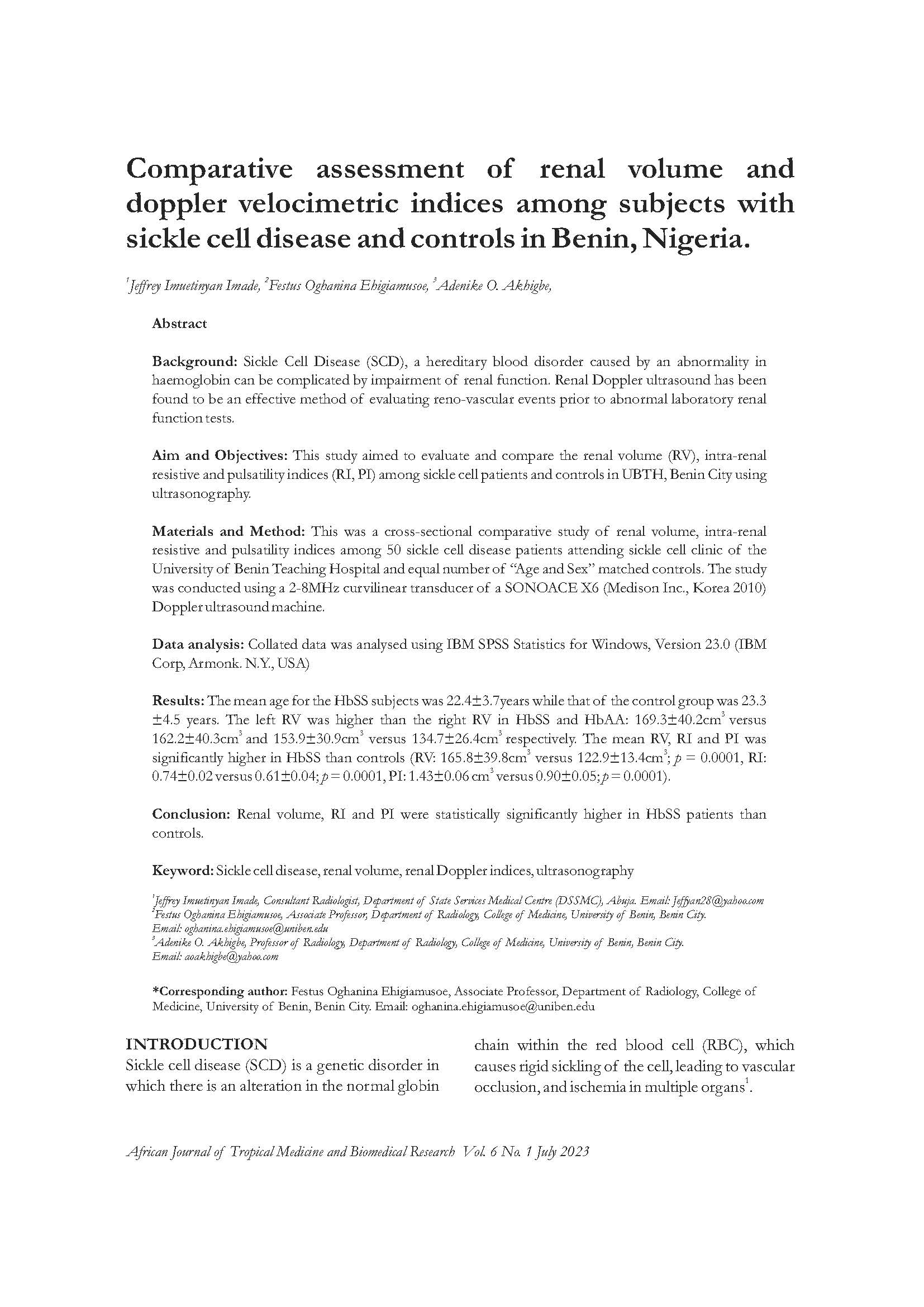
COMPARATIVE ASSESSMENT OF RENAL VOLUME AND DOPPLER VELOCIMETRIC INDICES AMONG SUBJECTS WITH SICKLE CELL DISEASE AND CONTROLS IN BENIN, NIGERIA.
Keywords:
Sickle cell disease, , renal volume, renal Doppler indices, ultrasonographyAbstract
Background: Sickle Cell Disease (SCD), a hereditary blood disorder caused by an abnormality in haemoglobin can be complicated by impairment of renal function. Renal Doppler ultrasound has been found to be an effective method of evaluating reno-vascular events prior to abnormal laboratory renal function tests.
Aim and Objectives: This study aimed to evaluate and compare the renal volume (RV), intra-renal resistive and pulsatility indices (RI, PI) among sickle cell patients and controls in UBTH, Benin City using
ultrasonography.
Materials and Method: This was a cross-sectional comparative study of renal volume, intra-renal resistive and pulsatility indices among 50 sickle cell disease patients attending sickle cell clinic of the University of Benin Teaching Hospital and equal number of “Age and Sex” matched controls. The study was conducted using a 2-8MHz curvilinear transducer of a SONOACE X6 (Medison Inc., Korea 2010) Doppler ultrasound machine.
Data analysis: Collated data was analysed using IBM SPSS Statistics for Windows, Version 23.0 (IBM
Corp, Armonk. N.Y., USA)
Results: The mean age for the HbSS subjects was 22.4±3.7 years while that of the control group was 23.3 ±4.5 years. The left RV was higher than the right RV in HbSS and HbAA: 169.3 ±40.2cm3 versus 162.2 ±40.3cm3 and 153.9 ±30.9cm3 versus 134.7 ±26.4cm3 respectively. The mean RV, RI and PI was significantly higher in HbSS than controls (RV: 165.8 ±39.8cm3 versus 122.9 ±13.4cm3 ; p = 0.0001, RI: 0.74±0.02 versus 0.61±0.04; p = 0.0001, PI: 1.43±0.06 cm3 versus 0.90±0.05; p = 0.0001).
Conclusion: Renal volume, RI and PI were statistically significantly higher in HbSS patients than controls.
References
Ma’aji SM, Jiya NM, Saidu SA,s Danfulani M, Yunusa GH, Sani UM, et al. Transabdominal sonographic findings in children with sickle cell anemia in Sokoto, North- eastern Nigeria. Niger J Basic Clin Sci 2012; 9: 14-7.
Herrick JB. Perculiar enlongated and sickle – shaped red blood corpuscles in a case of severe anemia. Arch Int Med 1910; 6:517- 520.
Bunn HF. Induction of fetal hemoglobin in sickle cell disease. Blood 1999; 93: 1787- 1789.
Piel FB, Hey SI, Gupta S, Weatherall DJ, Williams TN. Global burden of sickle cell anemia in children under five, 2010 -250:Modelling based on demographics, excess mortality, and interventions, PLos Med 2013; 10:e1001484.
Odame I. Perspective: we need a global solution. Nature. 2014; 515(7526):S10.
Tshilolo L, Kafando E, Sawadogo M, Cotton F, Vertongen F, Ferster A, et al. Neonatal screening and clinical care programmes for sickle cell disorders in Sub – Saharan Africa: lessons from pilot studies. Public Health. 2008 Sep; 122(9):933-41.
Kadima BT, Gini Ehungu JL, Ngiyulu RM, Ekulu PM, Aloni MN. High rate of sickle cell anemia in Sub- Saharan Africa underlines the need to screen all children with severe anemia for the disease. Acta Paediatr. 2015 Dec; 104(12): 1269-73.
Fleming AF, Storey J, Molineaux L, Iroko EA, Attai ED. Abnormal hemoglobin in the Sudan savanna of Nigeria. I. Prevalence of Hemoglobins and relationships between sickle cell trait, malaria and survival. Ann Trop Med and Parasitol. 1979 Apr;73(2): 161-72.
Uzoegwu PN, Onwurah AE Prevalence of hemoglobinopathy and Malaria Diseases in the Population of Old Aguata Division, Anambra State, Nigeria. Biokemistri. 2003;15(2):57-66.
Baba PI, Yvonne D, Juliana OI, John D, Matthew CE, Sukhleen MS, et al. Sickle Cell Disease Screening in Northern Nigeria: The Co-Existence of B-Thalassemia Inheritance. Pediat Therapeut 2015 Sept;5(3):1-4.
Odunvbun ME, Okolo AA, Rahimy CM. New born screening for sickle cell disease in a Nigerian Hospital. Public Health. 2008 Oct; 122(10):1111-6.
Nwogoh B, Adewoyin AS, Iheanacho OE, Bazuaye GN. “Prevalence of hemoglobin variants in Benin City, Nigeria.’’ Annals of Biomedical Sciences 2012; 11(2): 60-64.
Karl AN, Robert PH. Sickle Cell Disease: Renal Manifestations and Mechanisms. Nat Rev Nephrol.2015Mar;11(3):161-171.
Hebbel RP. Beyond Hemoglobin Polymerization: The Red Cell Membrane and Sickle Cell Disease Pathophysiology. Blood 1991 Jan;77(2):214-237.
Hebbel RP. Perspective series: Cell adhesion in vascular biology. Adhesive interactions of sickle erythrocytes and endothelium. J Clin Invest.1997Jun;99(11):2561-4.
Francis RB Jr, Johnson CS. Vascular occlusion in sickle cell disease: current concepts and unanswered questions. Blood,1991Apr;77(7):1405-1414.
Atalabi OM, Oyibotha OO, Akinola RA. Comparative analysis of Renal Dopppler indices in type 2 diabetes patients and healthy subjects in south western Nigeria. West African journal of ultrasound 2016; 17:12.
Atalabi OM, Yusuf BP. Comparative ultasound evaluation of renal resistive index in hypertensive and normotensive adults in Ibadan, south-west Nigeria. Trop. J Nephrol. 2012; (7): 19-26.
Tublin ME, Bude RO, Platt JF. The resistive Index in Renal Doppler Sonography: Where Do We Stand? AJR Am J Roentgenol. 2003 Apr; 180(4): 885-92.
Samir KB. More Definitions in Sickle Cell Disease: Steady State v Base line data. American Journal of Hematology 2011;87(3):338.
Rakhi PN, Carlton H Jr. Sickle cell trait diagnosis: clinical and social implications. Hematology Am Soc Hematol Educ Program 2015 Dec;2015(1):160-167.
Aikimbaev KS, Oguz M, Guvenc B, Baslamisli F, Kocak R. Spectral Pulsed Doppler Sonography of Renal vascular resistance in sickle cell disease: Clinical Implications. British Journal of Radiology 1996 Dec;69(828):1125-1129.
Ibinaiye PO, Babadoko AA, Hamidu AU, Hassan A, Yusuf R, Aiyekomogbon J, et al. Incidence of Abdominal Ultrasound Abnormalities in patients with sickle cell anemia in Zaria, Nigeria. European Journal of Scientific Research 2011;63 (4):548-556.
Shogbesan GA, Famurewa OC, Ayoola OO, Bolarinwa RA. Evaluation of Renal Artery Resistive and Pulsatility Index in steady state SCD patients and controls. West African Journal of Ultrasound 2017;18 (1):30-35.
Hamim AR. Abdominal Ultrasonographic Abnormalities in patients with Sickle Cell Anemia at Muhimbili National Hospital. M Med 2010 (Radiology) Dissertation, Muhimbili University of Health and Allied Sciences.
Kandrack MA, Graut KR, Segall A. Gender differences in Health-related behaviour: Some unanswered questions. Soc Sci Med 1991; 32: 579-90.
Cleary PD, Mechanic D Greenley IR. Sex differences in Medical Care Utilization: an empirical investigation. J Health Soc Behav. 1982; 23: 106-19.
Geofery L, Charles U, Muhammad S, Bukar AA, Ochie K. Sonographic Evaluation of Some Abdominal organs in Sickle Cell Disease in a Tertiary Health Institution in Northeastern Nigeria. J Med Ultrasound. 2018 Jan-Mar;26(1):31-36.
Rhodes M, Akohoue SA, Shankar SM, Fleming I et al. Growth pattern in children with sickle cell anaemia during puberty. Pediatr Blood Cancer. 2009 Oct;53(4):635-41.
Shilan HK, Naser AM, Ismaeel HA and Bryar AM. Comparative Ultrasonographic Measurement of Renal Size and its Correlation with Age, Gender, Body mass index in Normal Subjects in Sulaimani Region. European Scientific Journal. 2015; 11(12): 1857 – 7881.
Udoaka AI, Engi C and Agi CE. Sonological Evaluation of the liver, spleen and kidney in an adult southern Nigeria population. Asian Journal of Medical Science 2013; 5(2): 33 – 36.
Kanshal L, Verma VK, Ahirwar CP, Patil A, Singh SP, Sonographic Evaluation of Abdominal Organs in a sickle cell disease patient. International Journal of Medical Research and Review.2014 May; 2(3):202-207.
Balci A, Karazincir S, Sangun O, Gali E, Daplan T, Cingiz C et al. Prevalence of abdominal ultrasonographic abnormalities in patients with sickle cell disease. Diagn Interv Radiol 2008 Sept; 14(3):133-7.
Mohamed AE, Moawia G, Elsapi AA, Mahmoud SB, Mohamed E, Suliman S, et al. The Sonographic Assessment of Kidneys in patients with Sickle Cell Disease. Current Trends in Clinical and Medical Imaging 2017 Feb;1(1):001-004.
Nosiba HMA, Ahmed AM, Ala MAE. Evaluation of Abdominal Organs in Children with Sickle Cell Anemia Using Ultrasonography. Sch J App Med Sci 2017; 5(3C):920-925.
Ibinaiye PO, Aliyu AB, Rasheed Y, Abdul-Aziz H. Renal Complications of Sickle Cell Anemia in Zaria, Nigeria: An ultrasonographic assessment. West African Journal of Radiology 2013;20(1):19-22.
Rosado E, Paixao P, Schmitt W, Penha D, Carvalho FM, Tavares A, Amadora/PT. Sickle cell anemia- a review of imaging findings. European Society of Radiology (educational exhibit) 2014;C-1227:1-20.
Mapp E, Karasick S, Pollack H, Wechsler RJ, Karasick D. Uroradiological manifestations of S-hemoglobinopathy. Semin Roentgenol 1987 Jul; 22(3): 186 – 94.
Bhushita BL, Bhushan NL, Bhavana BL. Sonographic Screening for Abdominal organ Involvement in Sickle Cell Anemia- A step towards better patient care. Journal of Krishna Institute of Medical Sciences University 2017 Apr; 6(2):40-9.
Walker TM, Beardsall K, Thomas PW, Serjeant GR. Renal length in sickle cell disease: observations from a cohort study. Clin Nephrol.1996 Dec;46(6):384-8.
Kishor BT, Ritus SC, Vinod A, Suresh D, Virender S, Prashant N, et al. Renal Doppler Indices in Sickle Cell Disease: Early Radiologic Predictors of Renovascular changes. American Journal of Roentgenology 2008 Jul;191(1):239-242.
Sandra Regina Loggetto. Sickle cell anaemia: Clinical diversity and beta S-globin haplotypes. Rev Bras Hematol Hemoter.2013; 35 (3):153-62.
Mohamed AE, Mohamed EMG, Mohammed AA, Osman A, Elsafi AA. Impact of Sickle Cell Disease in Renal Arteries Blood flow Indices Using Ultrasonography. International Journal of Medical Imaging 2017;5 (2):9-13.
Bosma RJ, Ven der Heide JJ, Oosterop EJ, De Jong PE et al. Body Mass Index is associated with altered renal hemodynamics in non-obese healthy subjects. Kidney Int.2004 Jan;65(1):259-65.
Bachir D, Maurel A, Benzard Y, Razavian M, et al. Improvement of Microcirculation Abnormalities in Sickle Cell Patients upon Buflomedil Treatment. Microvas. Res. 1993; 46(3):359-373.

Downloads
Published
Issue
Section
License
Copyright (c) 2023 African Journal of Tropical Medicine and Biomedical Research

This work is licensed under a Creative Commons Attribution-ShareAlike 4.0 International License.
Key Terms:
- Attribution: You must give appropriate credit to the original creator.
- NonCommercial: You may not use the material for commercial purposes.
- ShareAlike: If you remix, transform, or build upon the material, you must distribute your contributions under the same license as the original.
- No additional restrictions: You may not apply legal terms or technological measures that legally restrict others from doing anything the license permits.
For full details, please review the Complete License Terms.


